Bassett Collection of Stereoscopic Images of Human Anatomy
Exploration of the meninges and brain in situ
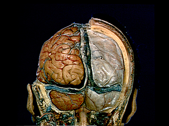
Posterior view of cranial meninges
Image #1-5
KEYWORDS: Bones cartilage joints, Brain, Cerebellum, Meninges, Occipital lobe, Scalp, Telencephalon, Overview.
Creative Commons
Stanford holds the copyright to the David L. Bassett anatomical images and has assigned Creative Commons license Attribution-Share Alike 4.0 International to all of the images.

For additional information regarding use and permissions, please contact the Medical History Center.

Exploration of the meninges and brain in situ
Posterior view of cranial meninges
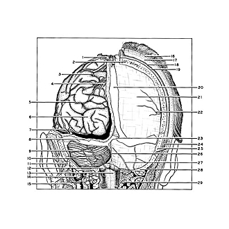
On the right the calvaria and layers of the scalp are shown in relation to the dura. On the left the dura has been cut away to reveal the cerebral hemisphere and cerebellum covered with the arachnoid membrane. The confluence of the sinuses (23) is shown.
- Venous lacuna
- Diploic vein within parietal bone
- Parieto-occipital branch of posterior cerebral artery
- Superior cerebral veins (posterior branches)
- Posterior parietal branch of middle cerebral artery
- Occipital lobe
- Inferior cerebral vein (lateral occipital vein)
- Tentorium cerebelli and transverse sinus (opened)
- Horizontal cerebellar sulcus
- Branch posterior inferior cerebellar artery (PICA)
- Mastoid emissary
- Cerebellar tonsil (at level of foramen magnum)
- Medulla viewed through arachnoid membrane
- Superior obliquus capitis muscle (cut across)
- Occipitalis vein (note posterior vertebral plexus more medial and deeper in the dissection)
- Skin
- Superficial fascia
- Galea aponeurotica
- Pericranium
- Superior sagittal sinus
- Dura mater
- Posterior branch of middle meningeal artery
- Confluence of the sinuses
- Occipitalis muscle (cut across)
- Temporal bone and occipitomastoid suture
- Occipital sinus
- Posterior meningeal artery
- Occipital bone
- Posterior arch of atlas (cut across)