Bassett Collection of Stereoscopic Images of Human Anatomy
Pectoral region and axilla
Superficial structures of left arm and thorax, anterior view
Image #89-1
KEYWORDS: Axilla, Fascia ligaments and tendons, Neuralnetwork, Pectoral region, Peripheral nervous system, Upper arm, Overview.
Creative Commons
Stanford holds the copyright to the David L. Bassett anatomical images and has assigned Creative Commons license Attribution-Share Alike 4.0 International to all of the images.

For additional information regarding use and permissions, please contact the Medical History Center.
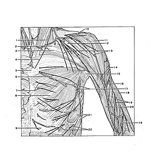
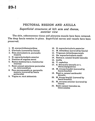
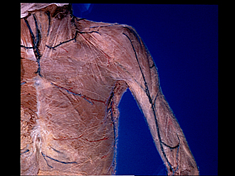
Pectoral region and axilla
Superficial structures of left arm and thorax, anterior view
The skin, subcutaneous tissue and platysma muscle have been removed. The deep fascia remains in place. Superficial nerves and vessels have been preserved.
- Sternocleidomastoid muscle
- Clavicle (covered by fascia)
- Clavicular part pectoralis major muscle
- Anterior supraclavicular nerve
- Position of sternal angle
- Mammary branch internal mammary artery
- Anterior pectoral cutaneous branches intercostal nerves
- Sternocostal part pectoralis major muscle (covered by pectoral fascia)
- Sheath of rectus abdominis muscle
- Posterior supraclavicular nerve
- Deltoid muscle (covered by fascia)
- Deltopectoral triangle
- Middle supraclavicular nerves
- Branch of lateral brachial cutaneous nerve
- Axilla
- Cephalic vein
- Lateral thoracic artery and thoracoepigastric vein
- Triceps brachii muscle (covered by brachial fascia)
- Branches of dorsal antebrachial cutaneous nerve
- Biceps brachii muscle (covered by brachial fascia)
- Serratus anterior muscle (covered by fascia)
- Lateral cutaneous branches of intercostal nerves