Bassett Collection of Stereoscopic Images of Human Anatomy
Dissection of anterior and lateral regions of neck
Relations of thyroid gland, anterior view
Image #74-7
KEYWORDS: Exocrine and endocrine, Peripheral nervous system, Throat, Vasculature.
Creative Commons
Stanford holds the copyright to the David L. Bassett anatomical images and has assigned Creative Commons license Attribution-Share Alike 4.0 International to all of the images.

For additional information regarding use and permissions, please contact the Medical History Center.
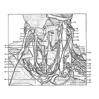
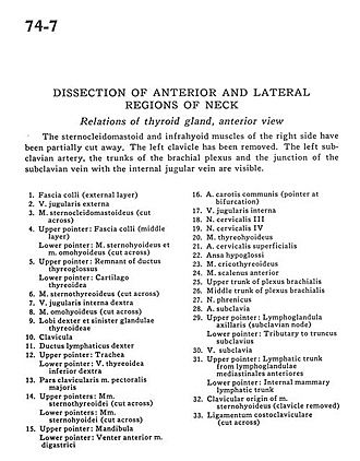
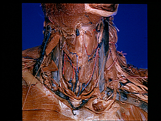
Dissection of anterior and lateral regions of neck
Relations of thyroid gland, anterior view
The sternocleidomastoid and infrahyoid muscles of the right side have been partially cut away. The left clavicle has been removed. The left subclavian artery, the trunks of the brachial plexus and the junction of the subclavian vein with the internal jugular vein are visible.
- Superficial fascia (external layer)
- External jugular vein
- Sternocleidomastoid muscle (cut across)
- Upper pointer: Superficial fascia (middle layer) Lower pointer: Sternohyoid muscle and omohyoid muscle (cut across)
- Upper pointer: Remnant of thyroglossal duct Lower pointer: Thyroid cartilage
- Sternothyroid muscle (cut across)
- Internal jugular vein right
- Omohyoid muscle (cut across)
- Right and left lobe of thyroid gland
- Clavicle
- Right lymphatic duct
- Upper pointer: Trachea Lower pointer: Inferior thyroid vein right
- Clavicular part pectoralis major muscle
- Upper pointers: Sternothyroid muscles (cut across) Lower pointers: Sternohyoid muscles (cut across)
- Upper pointer: Mandible Lower pointer: Anterior belly digastric. muscle
- Common carotid artery (pointer at bifurcation)
- Internal jugular vein
- Cervical nerve III
- Cervical nerve IV
- Thyrohyoid muscle
- Superficial cervical artery
- Ansa hypoglossi
- Cricothyroid muscle
- Anterior scalene muscle
- Upper trunk of brachial plexus
- Middle trunk of brachial plexus
- Phrenic nerve
- Subclavian artery
- Upper pointer: Axillary lymph node (subclavian node) Lower pointer: Tributary to subclavian trunk
- Subclavian vein
- Upper pointer: Lymphatic trunk from anterior mediastinal lymph nodes Lower pointer: Internal mammary lymphatic trunk
- Clavicular origin of sternohyoid muscle (clavicle removed)
- Costoclavicular ligament (cut across)