Bassett Collection of Stereoscopic Images of Human Anatomy
Dissection of anterior and lateral regions of neck
Thyrohyoid muscle, anterior view
Image #74-3
KEYWORDS: Muscles and tendons, Throat.
Creative Commons
Stanford holds the copyright to the David L. Bassett anatomical images and has assigned Creative Commons license Attribution-Share Alike 4.0 International to all of the images.

For additional information regarding use and permissions, please contact the Medical History Center.
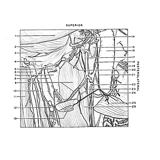
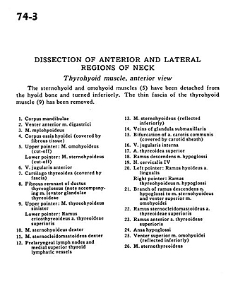
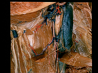
Dissection of anterior and lateral regions of neck
Thyrohyoid muscle, anterior view
The sternohyoid and omohyoid muscles (5) have been detached from the hyoid bone and turned inferiorly. The thin fascia of the thyrohyoid muscle (9) has been removed.
- Body of mandible
- Anterior belly digastric muscle
- Mylohyoid muscle
- Body hyoid bone (covered by fibrous tissue)
- Upper pointer: Omohyoid muscle (cut-off) Lower pointer: Sternohyoid muscle (cut-off)
- Anterior jugular vein
- Thyroid cartilage (covered by fascia)
- Fibrous remnant of thyroglossal duct (note accompanying Ievator glandulae thyroideae muscle)
- Upper pointer: Thyrohyoid muscle left Lower pointer: Cricothyroid branch superior thyroid artery
- Sternohyoid muscle right
- Sternocleidomastoid muscle right
- Prelaryngeal lymph nodes and medial superior thyroid lymphatic vessels
- Sternohyoid muscle (reflected inferiorly)
- Veins of submandibular gland
- Bifurcation of common carotid artery (covered by carotid sheath)
- Internal jugular vein
- Superior thyroid artery
- Descending branch hypoglossal nerve
- Cervical nerve IV
- Left pointer: Hyoid branch lingual artery Right pointer: Thyrohyoid branch hypoglossal nerve
- Branch of descending branch hypoglossal nerve to sternohyoid muscle and superior belly omohyoid muscle
- Sternocleidomastoid branch superior thyroid artery
- Anterior branch of superior thyroid artery
- Ansa hypoglossi
- Superior belly omohyoid muscle (reflected inferiorly)
- Sternothyroid muscle