Bassett Collection of Stereoscopic Images of Human Anatomy
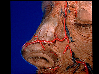
Dissection of nose
External nasal nerve; arteries of nose, left anterolateral view
Image #71-1
KEYWORDS: Bones cartilage joints, Connective tissue, Face, Nose, Peripheral nervous system, Vasculature.
Creative Commons
Stanford holds the copyright to the David L. Bassett anatomical images and has assigned Creative Commons license Attribution-Share Alike 4.0 International to all of the images.

For additional information regarding use and permissions, please contact the Medical History Center.
Dissection of nose
External nasal nerve; arteries of nose, left anterolateral view
The transverse part of the nasal muscle has been removed. Two branches (6,8) of the external nasal nerve are visible. These emerge through the membrane which covers the junction of the nasal bone and lateral nasal cartilage. A filament of the external nasal branch (10) of the infraorbital nerve crosses the external nasal nerves superficially. No communication was found between the nerves.
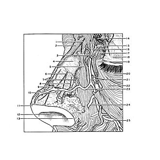
- Branch infratrochlear nerve
- Procerus muscle
- Dorsal nasal artery
- Periosteum of nasal bone
- Angular head of levator labii superioris muscle
- External nasal branch of anterior ethmoidal nerve (medial branch)
- Transverse part of nasalis muscle
- External nasal branch of anterior ethmoidal nerve (lateral branch)
- Perichondrium of lateral nasal cartilage
- External nasal branch of infraorbital nerve
- Apex of nose
- Naris
- Ala nasi
- Infratrochlear nerve
- Middle palpebral artery
- Frontal artery
- Orbicularis oculi muscle (cut across)
- Superior tarsus
- Medial palpebral ligament
- Palpebral part of orbicularis oculi muscle
- Angularis artery and vein
- Orbital part orbicularis oculi muscle
- Nasal branch of angular artery
- Alar branch of angular artery
- Infraorbital head of levator labii superioris muscle