
Bassett Collection of Stereoscopic Images of Human Anatomy
Microradiograph of eye; central optic pathways and related structures
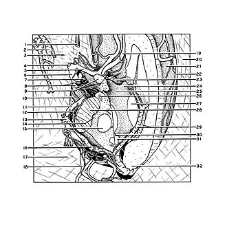
Relations of optic pathways at base of brain.
Image #58A-5
KEYWORDS: Brain, Diencephalon, Eye, Face, Midbrain, Peripheral nervous system, Vasculature.
Creative Commons
Stanford holds the copyright to the David L. Bassett anatomical images and has assigned Creative Commons license Attribution-Share Alike 4.0 International to all of the images.

For additional information regarding use and permissions, please contact the Medical History Center.

Microradiograph of eye; central optic pathways and related structures
Relations of optic pathways at base of brain.
The specimen shown in the preceding photograph has been reoriented for this view so that the left orbit extends above and to the left of the area included in the photograph. The brain stem has been cut across through the rostral part of the mesencephalon. The left optic tract has been removed and the optic chiasm has been lifted slightly out of its normal position. The components of the arterial circle of Willis that are related to the optic pathways (i.e., internal carotid artery (6), anterior cerebral artery (5), anterior communicating artery (between 5 and 19, unlabeled), posterior communicating artery (8) and posterior cerebral artery (11)) remain in position.
- Olfactory tract (cut across)
- Inferior cerebral vein
- Ala parva ossis sphenoidalis (covered by dura)
- Medial cerebral artery (cut across)
- Left anterior cerebral artery
- Internal carotid artery
- Chiasmatic cistern
- Upper pointer: Anterior choroid artery (cut across) Lower pointer: Posterior communicating artery
- Optic tract (cut across and elevated)
- Oculomotor nerve (III)
- Posterior cerebral artery
- Central ramus of posterior cerebral artery
- Tributaries of the basal vein which passed into tip of inferior horn of lateral ventricle
- Substantia nigra
- Nucleus ruber
- Basal vein
- Cerebellar tentorium
- Continuation of main trunk of posterior cerebral artery
- Right anterior cerebral artery (anterior communicating artery partly obscured by optic chiasm)
- Falx cerebri
- Cingulate gyrus
- Pericallosal branch of anterior cerebral artery
- Anterior commissure
- Pars libera columnae fornicis
- Ventricular surface of hypothalamus
- Mamillary body (cut across)
- Intermediate mass
- Cerebral peduncle
- Internal cerebral veins
- Aditus ad aqueductum cerebri
- Posterior commissure
- Splenium corporis callosi
- [Legend above restored translation from Latin]