Bassett Collection of Stereoscopic Images of Human Anatomy
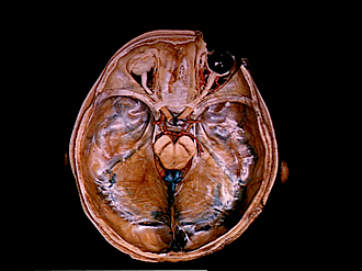
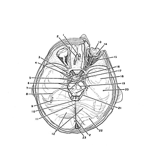
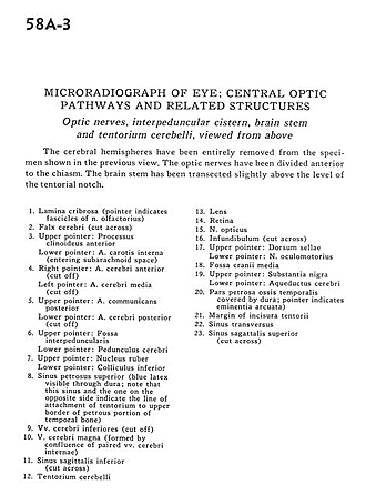
Microradiograph of eye; central optic pathways and related structures
Optic nerves, interpedicular cistern, brain stem and tentorium cerebelli, viewed from above
Image #58A-3
KEYWORDS: Brain, Diencephalon, Eye, Face, Midbrain, Occipital lobe, Peripheral nervous system, Telencephalon.
Creative Commons
Stanford holds the copyright to the David L. Bassett anatomical images and has assigned Creative Commons license Attribution-Share Alike 4.0 International to all of the images.

For additional information regarding use and permissions, please contact the Medical History Center.
Microradiograph of eye; central optic pathways and related structures
Optic nerves, interpedicular cistern, brain stem and tentorium cerebelli, viewed from above
The cerebral hemispheres have been entirely removed from the specimen shown in the previous view. The optic nerves have been divided anterior to the chiasm. The brain stem has been transected slightly above the level of the tentorial notch.
- Lamina cribrosa (pointer indicates fascicles of olfactory nerve)
- Falx cerebri (cut across)
- Upper pointer: Anterior clinoid process Lower pointer: Internal carotid artery (entering subarachnoid space)
- Right pointer: Anterior cerebral artery (cut off) Left pointer: Medial cerebral artery (cut off)
- Upper pointer: Posterior communicating artery Lower pointer: Posterior cerebral artery (cut off)
- Upper pointer: Interpeduncular fossa Lower pointer: Cerebral peduncle
- Upper pointer: Nucleus ruber Lower pointer: Colliculus inferior
- Superior petrosal sinus (blue latex visible through dura; note that this sinus and the one on the opposite side indicate the line of attachment of tentorium to upper border of petrous portion of temporal bone)
- Inferior cerebral veins (cut off)
- Great cerebral vein (formed by confluence of paired internal cerebral veins)
- Inferior sagittal sinus (cut across)
- Cerebellar tentorium
- Lens
- Retina
- Optic nerve
- Infundibulum (cut across)
- Upper pointer: Dorsum sellae Lower pointer: Oculomotor nerve
- Medial cranial fossa
- Upper pointer: Substantia nigra Lower pointer: Cerebral aqueduct
- Petrosal part of the temporal bone (covered by dura; pointer indicates eminentia arcuata)
- Margin of the tentorial incisure
- Transverse sinus
- Superior sagittal sinus (cut across)
- [Legend above restored translation from Latin]