Bassett Collection of Stereoscopic Images of Human Anatomy
Orbital fascia
Fascia related to left eyeball, posterior view
Image #57-2
KEYWORDS: Connective tissue, Eye, Face.
Creative Commons
Stanford holds the copyright to the David L. Bassett anatomical images and has assigned Creative Commons license Attribution-Share Alike 4.0 International to all of the images.

For additional information regarding use and permissions, please contact the Medical History Center.
Orbital fascia
Fascia related to left eyeball, posterior view
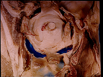
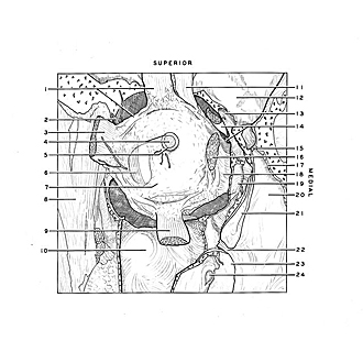
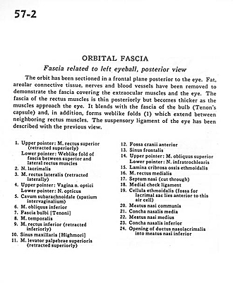
The orbit has been sectioned in a frontal plane posterior to the eye. Fat, areolar connective tissue, nerves and blood vessels have been removed to demonstrate the fascia covering the extraordinary muscles and the eye. The fascia of the rectus muscles is thin posteriorly but becomes thick as the muscles approach the eye. It blends with the fascia of the bulb (Tenon's capsule) and, in addition, forms weblike folds (1) which extend between neighboring rectus muscles. The suspensory ligament of the eye has been described with the previous view.
- Upper pointer: Superior rectus muscle (retracted superiorly) Lower pointer: Weblike fold of fascia between superior and lateral rectus muscles
- Lacrimal nerve
- Lateral rectus muscle (retracted laterally)
- Upper pointer: Sheath of optic nerve Lower pointer: Optic nerve
- Subarachnoid space (spatium intervaginalium)
- Inferior oblique muscle
- Bulbar fascia
- Temporalis muscle
- Inferior rectus muscle (retracted inferiorly)
- Maxillary sinus
- Levator palpebrae superioris muscle (retracted superiorly)
- Anterior cranial fossa
- Frontal sinus
- Upper pointer: Superior oblique muscle Lower pointer: Infratrochlear nerve
- Cribriform plate ethmoid bone
- Medial rectus muscle
- Nasal septum (cut through)
- Medial check ligament
- Ethmoidal cell (fossa for lacrimal sac lies anterior to this air cell)
- Common nasal meatus
- Middle nasal concha
- Middle nasal meatus
- Inferior nasal concha
- Opening of nasolacrimal duct into Inferior nasal meatus