
Bassett Collection of Stereoscopic Images of Human Anatomy
General orientation views of dissection
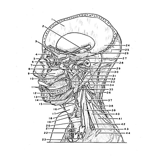
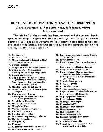
Deep dissection of head and neck, left lateral view; brain removed
Image #49-7
KEYWORDS: Brain, Cheek, Connective tissue, Face, Midbrain, Muscles and tendons, Peripheral nervous system, Vasculature, Fascia and connective tissue, Overview.
Creative Commons
Stanford holds the copyright to the David L. Bassett anatomical images and has assigned Creative Commons license Attribution-Share Alike 4.0 International to all of the images.

For additional information regarding use and permissions, please contact the Medical History Center.
General orientation views of dissection
Deep dissection of head and neck, left lateral view; brain removed
The left half of the calvaria has been removed and the cerebral hemispheres cut away to expose the left optic tract (2) encircling the cerebral peduncle (26). The close-up views which illustrate more details of this dissection are to be found as follows
- Falx cerebri
- Optic tract
- Lateral rectus muscle (lateral wall of orbit cut away)
- Superior tarsus
- Ophthalmic nerve in cavernous sinus
- Upper pointer: Maxillary nerve Lower pointer: Sphenopalatine artery
- Nasal cavity (opened)
- Upper pointer: Buccal nerve (crossing internal maxillary artery) Lower pointer: Lateral plate of pterygoid process
- Maxilla (partially cut away)
- Buccinator muscle (cut away to expose tongue)
- Upper pointer: Tongue Lower pointer: Medial incisor (sectioned)
- Sublingual gland
- Mandible (cut across)
- Mylohyoid muscle
- Anterior belly digastric muscle
- Common carotid artery
- Thyroid cartilage
- Inferior pharyngeal constrictor muscle
- Cricothyroid muscle
- Vagus nerve (X)
- Trachea
- Common carotid artery (cut across)
- Sternoclavicular articulation
- Junction of tentorium cerebelli with falx cerebri
- Lambdoidal suture
- Upper pointer: Lateral geniculate body Lower pointer: Base of peduncles
- Semilunar ganglion (CN V)
- Upper pointer: External acoustic meatus (auricle removed) Lower pointer: Mastoid cells (dissected)
- Auriculotemporal nerve
- Facial nerve (VII)
- External carotid artery
- Posterior belly of digastric muscle
- Upper pointer: Inferior alveolar nerve Lower pointer: Lingual nerve
- Internal pterygoid muscle
- Hypoglossal nerve (XII)
- Longus capitis muscle
- Cervical nerve IV
- Longissimus cervicis muscle
- Anterior scalene muscle
- Middle scalene muscle
- Posterior scalene muscle
- Brachial plexus (cut across through roots)
- Subclavian artery (cut off)
- Rib I (periosteum removed to show small area of rib)