Bassett Collection of Stereoscopic Images of Human Anatomy
Osteology
Lateral wall of right nasal fossa
Image #38-1
KEYWORDS: Bones cartilage joints, Face, Nose.
Creative Commons
Stanford holds the copyright to the David L. Bassett anatomical images and has assigned Creative Commons license Attribution-Share Alike 4.0 International to all of the images.

For additional information regarding use and permissions, please contact the Medical History Center.
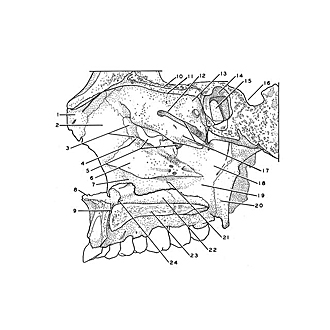
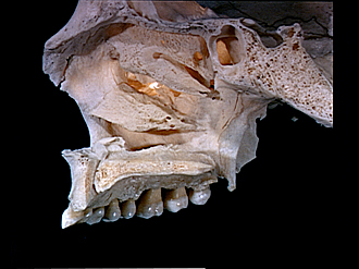
Osteology
Lateral wall of right nasal fossa
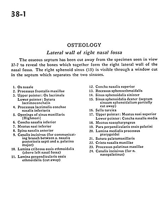
The osseous septum has been cut away from the specimen seen in view 37-7 to reveal the bones which together form the right lateral wall of the nasal fossa. The right sphenoid sinus (15) is visible through a window cut in the septum which separates the two sinuses.
- Nasal bone
- Frontal process of maxilla
- Upper pointer: Lacrimal bone Lower pointer: Lacrimoconchal suture
- Lacrimal process inferior nasal conchae
- Openings of maxillary sinus
- Inferior nasal concha
- Inferior nasal meatus
- Anterior nasal spine
- Incisive canal (for communicating branch between posterior nasal septal artery and greater palatine artery)
- Cribriform plate éthmoid bone (above left nasal fossa)
- Perpendicular plate of ethmoid bone (cut away)
- Superior nasal concha
- Sphenoethmoidal recess
- Sphenoid sinus left
- Sphenoid sinus right (sphenoidal septal sinuses partially cut away)
- Sella turcica
- Upper pointer: Superior nasal meatus Lower pointer: Middle nasal concha
- Nasopharyngeal meatus
- Perpendicular part of palatine bone
- Medial plate of pterygoid process
- Palatomaxillary suture
- Nasomaxillary crest
- Palatine process of maxilla
- Incisive canal (for nasopalatine nerve)