Bassett Collection of Stereoscopic Images of Human Anatomy
Dissection of anterior aspect of vertebral column
Sacral region.
Image #221-5
KEYWORDS: Sacral region, Vasculature, Vertebral column.
Creative Commons
Stanford holds the copyright to the David L. Bassett anatomical images and has assigned Creative Commons license Attribution-Share Alike 4.0 International to all of the images.

For additional information regarding use and permissions, please contact the Medical History Center.
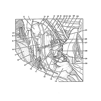
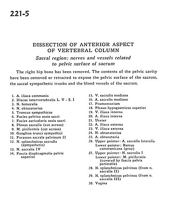
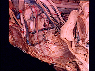
Dissection of anterior aspect of vertebral column
Sacral region.
The right hip bone has been removed. The contents of the pelvic cavity have been removed or retracted to expose the pelvic surface of the sacrum, the sacral sympathetic trunks and the blood vessels of the sacrum
- Common iliac artery
- Intervertebral disc L. V - S. I
- Femoral nerve
- Obturator nerve
- Sympathetic trunk
- Pelvic surface sacrum
- Articular surface sacrum
- Sacral plexus (cut across)
- Piriform muscle (cut across)
- Ganglion of sympathetic trunk
- Pelvic sacral foramen II
- Sacral splanchnic nerve (sympathetic)
- Sacral nerve IV
- Superior diaphragmatic pelvic fascia
- Median sacral vein
- Median sacral artery
- Promontory
- Superior hypogastric plexus
- Internal iliac vein
- Internal iliac artery
- Ureter
- External iliac artery
- External iliac vein
- Obturator nerve
- Obturator artery
- Upper pointer: Lateral sacral artery Lower pointer: Ramus communicans (gray)
- Upper pointer: Sacral nerve I Lower pointer: Piriform muscle (covered by parietal pelvic fascia)
- Pelvic splanchnic nerve (from sacral nerve II)
- Pelvic splanchnic nerve (from sacral nerve III) 30. Vagina