
Bassett Collection of Stereoscopic Images of Human Anatomy
Exploration of the meninges and brain in situ
Olfactory bulb and tract in situ
Image #2-5
KEYWORDS: Brain, Peripheral nervous system, Telencephalon.
Creative Commons
Stanford holds the copyright to the David L. Bassett anatomical images and has assigned Creative Commons license Attribution-Share Alike 4.0 International to all of the images.

For additional information regarding use and permissions, please contact the Medical History Center.

Exploration of the meninges and brain in situ
Olfactory bulb and tract in situ
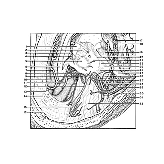
The view is from above and in front. A cut has been made through the basal ganglia close to the attachment of the olfactory tract to the base of the brain, and the left hemisphere removed. The olfactory bulb and tract remain in position on the floor of the anterior cranial fossa. The most medial part of the lateral fissure is opened from above and its contained vessels (28) exposed. The temporal pole is still present.
- Caudate nucleus (cut across)
- Internal capsule (cut across)
- Fornix (column) (cut across)
- Interventricular foramen (of Monro)
- Globus pallidus
- Corpus callosum
- Pericallosal branch of anterior cerebral artery
- Medial olfactory stria
- Optic nerve (11)
- Cingulate gyrus
- Anterior cerebral artery left (cut off)
- Olfactory tract 29.
- Callosomarginal branch of anterior cerebral artery right
- Falx cerebri
- Olfactory bulb
- Superior sagittal sinus (opened)
- Choroid plexus lateral ventricle
- Inferior longitudinal fasciculus
- Insula
- External capsule
- Putamen
- Claustrum
- Medullary substance of insula
- "Substantia innominata" and uncus hippocampal gyrus
- Superior temporal gyrus (cut across)
- Uncinate fasciculus (cut across)
- Lateral olfactory stria
- Middle cerebral artery
- Middle cerebral vein
- Dura mater (note: cut ends of middle meningeal vessels near pointer)
- Temporalis muscle (cut across)
- Orbital part of frontal bone (covered with dura and arachnoid)