Bassett Collection of Stereoscopic Images of Human Anatomy
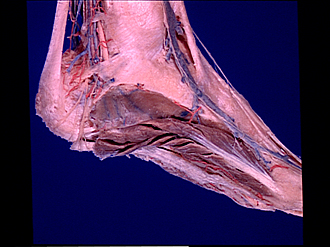
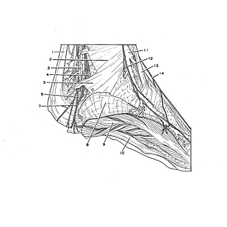
Dissection of medial aspect of left foot and ankle
Abductor hallucis reflected; posterior tibial vessels and tibial nerve at ankle, medial view
Image #198-7
KEYWORDS: Ankle, Foot and toes, Muscles and tendons, Peripheral nervous system, Vasculature.
Creative Commons
Stanford holds the copyright to the David L. Bassett anatomical images and has assigned Creative Commons license Attribution-Share Alike 4.0 International to all of the images.

For additional information regarding use and permissions, please contact the Medical History Center.
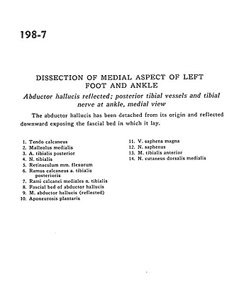
Dissection of medial aspect of left foot and ankle
Abductor hallucis reflected; posterior tibial vessels and tibial nerve at ankle, medial view
The abductor hallucis has been detached from its origin and reflected downward exposing the fascial bed in which it lay.
- Tendo calcaneus
- Medial malleolus
- Posterior tibial artery
- Tibial nerve
- Flexor retinaculum
- Calcaneal branch of the posterior tibial artery
- Medial calcaneal branches of tibial nerve
- Fascial bed of abductor hallucis
- Abductor hallucis muscle (reflected)
- Plantar aponeurosis
- Great saphenous vein
- Saphenous nerve
- Tibialis anterior muscle
- Dorsal medial cutaneous nerve