Bassett Collection of Stereoscopic Images of Human Anatomy
Exploration of those parts of the brain supplied by the posterior cerebral artery
Cingulum and inferior longitudinal fasciculus (continued)
Image #19-4
KEYWORDS: Brain, Telencephalon, Vasculature.
Creative Commons
Stanford holds the copyright to the David L. Bassett anatomical images and has assigned Creative Commons license Attribution-Share Alike 4.0 International to all of the images.

For additional information regarding use and permissions, please contact the Medical History Center.
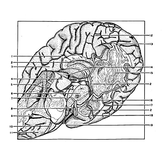
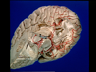
Exploration of those parts of the brain supplied by the posterior cerebral artery
Cingulum and inferior longitudinal fasciculus (continued)
The gyrus cinguli has been partially removed to reveal the course of the cingulum above the corpus callosum. Cortex which lay deep in the collateral fissure has been scraped away and the fusiform gyrus partly removed. More of the inferior longitudinal fasciculus is thus exposed. The brain is now turned so that more of the basal surface is shown than previously.
- Cingulate sulcus
- Cingulum
- Cingulate gyrus (partly resected)
- Pulvinar
- Internal capsule (cut across)
- Mesencephalon (cut across)
- Posterior cerebral artery left
- Basilar artery (cut across)
- Middle cerebral artery left (cut across)
- Olfactory tract
- Inferior surface of left frontal lobe
- Parieto-occipital fissure
- Calcarine fissure
- Corpus callosum (divided, left half mostly removed)
- Inferior longitudinal fasciculus
- Posterior cerebral artery right
- Inferior temporal sulcus
- Collateral fissure
- Temporal pole