Bassett Collection of Stereoscopic Images of Human Anatomy
Radiography
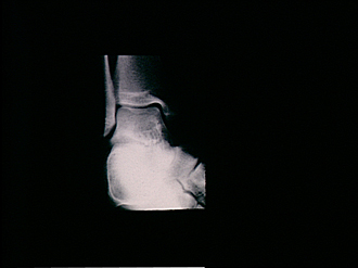
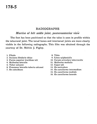
Mortise of left ankle joint, posteroanterior view
Image #178-5
KEYWORDS: Ankle, Bones joints cartilage.
Creative Commons
Stanford holds the copyright to the David L. Bassett anatomical images and has assigned Creative Commons license Attribution-Share Alike 4.0 International to all of the images.

For additional information regarding use and permissions, please contact the Medical History Center.
Radiography
Mortise of left ankle joint, posteroanterior view
The foot has been positioned so that the talus is seen in profile within the talocrural joint. The tarsal bones and intertarsal joints are more clearly visible in the following radiographs. This film was obtained through the courtesy of Dr. Melvin J. Figley.
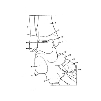
- Fibula
- Fibular notch
- Superior trochlear surface of talus
- Lateral malleolus
- Tuberosity of calcaneus
- Lateral tubercle process of calcaneus
- Cuboid bone
- Tibia
- Epiphysial line
- Talocrural articular space
- Medial malleolus
- Head of talus
- Navicular bone
- Intermediate cuneiform bone
- Medial cuneiform bone
- Lateral cuneiform bone