
Bassett Collection of Stereoscopic Images of Human Anatomy
Embryo, placenta and fetal membranes
Three-month-old fetus in situ within fetal membranes
Image #164-6
KEYWORDS: Uterus, Muscles and tendons.
Creative Commons
Stanford holds the copyright to the David L. Bassett anatomical images and has assigned Creative Commons license Attribution-Share Alike 4.0 International to all of the images.

For additional information regarding use and permissions, please contact the Medical History Center.

Embryo, placenta and fetal membranes
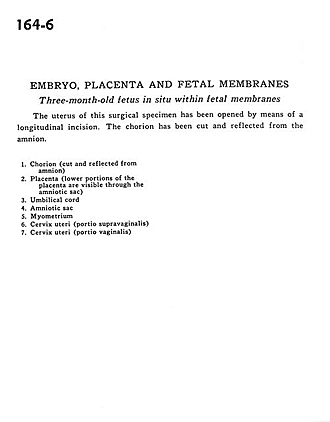
Three-month-old fetus in situ within fetal membranes
The uterus of this surgical specimen has been opened by means of a longitudinal incision. The chorion has been cut and reflected from the amnion.
- Chorion (cut and reflected from amnion)
- Placenta (lower portions of the placenta are visible through the amniotic sac)
- Umbilical cord
- Amniotic sac
- Myometrium
- Cervix of uterus (supravaginal part)
- Cervix of uterus (vaginal part)