
Bassett Collection of Stereoscopic Images of Human Anatomy
Exploration of the brain from the medial aspect
Stria medullaris thalami; mammillothalamic tract entering anterior nucleus of thalamus; thalamic branches of posterior cerebral artery
Image #16-5
KEYWORDS: Brain, Diencephalon, Midbrain, Vasculature.
Creative Commons
Stanford holds the copyright to the David L. Bassett anatomical images and has assigned Creative Commons license Attribution-Share Alike 4.0 International to all of the images.

For additional information regarding use and permissions, please contact the Medical History Center.
Exploration of the brain from the medial aspect
Stria medullaris thalami; mammillothalamic tract entering anterior nucleus of thalamus; thalamic branches of posterior cerebral artery
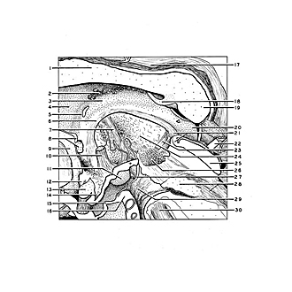
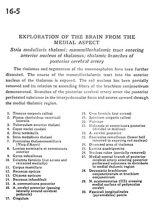
The thalamus and tegmentum of the mesencephalon have been further dissected. The course of the mammillothalamic tract into the anterior nucleus of the thalamus is exposed. The red nucleus has been partially removed and its relation to ascending fibers of the brachium conjunctivum demonstrated. Branches of the posterior cerebral artery enter the posterior perforated substance in the interpeduncular fossa and course upward through the medial thalamic region.
- Corpus callosum (trunk)
- Choroid plexus lateral ventricle
- Anterior tubercle of thalamus
- Head of caudate nucleus
- Stria terminalis
- Stria medullaris thalami
- Mamillothalamic tract
- Lamina terminalis and anterior commissure
- Subcallosal gyrus
- Fornix (column) (cut across and retracted medially)
- Mamillary body
- Optic recess
- Optic chiasm
- Infundibular recess
- Posterior communicating artery
- Posterior cerebral artery (passing laterally around cerebral peduncle)
- Cingulum
- Fornix (crus) (cut across)
- Corpus callosum (splenium)
- Pulvinar
- Habenula and posterior commissure (divided in midline)
- Posterior cerebral artery
- Fasciculus retroflexus (lower half removed in exposing red nucleus)
- Dissected area of thalamus
- Quadrigeminal plate
- Red nucleus (partially removed)
- Medial central branch of posterior cerebral artery entering posterior perforated substance to distribute to medial thalamic region
- Decussation brachium conjunctivum (superior cerebellar peduncle) and brachium conjunctivum (superior cerebellar peduncle)
- Oculomotor nerve (III) crossing medial surface of cerebral peduncle
- Longitudinal fasciculus (pyramidal)