Bassett Collection of Stereoscopic Images of Human Anatomy
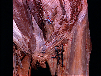
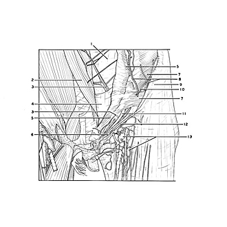
Dissection of female inguinal region
Inguinal canal (continued).
Image #137-6
KEYWORDS: Muscles and tendons, Peripheral nervous system.
Creative Commons
Stanford holds the copyright to the David L. Bassett anatomical images and has assigned Creative Commons license Attribution-Share Alike 4.0 International to all of the images.

For additional information regarding use and permissions, please contact the Medical History Center.
Dissection of female inguinal region
Inguinal canal (continued).
The lower abdominal wall has been dissected more completely so that relations of the inguinal canal to other structures of the abdominal wall are evident. The anterior lamina of the rectus sheath has been resected and the rectus muscle has been reflected medially. Lateral to the rectus sheath the transversus abdominis muscle (7) remains intact and short ends of the divided internal oblique (5) are visible. Structures in the inguinal canal remain essentially unchanged from the situation shown in the previous view (137-5)
- Sheath of rectus abdominis muscle (posterior layer, semicircular or arcuate line not distinct)
- Rectus abdominis muscle (reflected medially)
- Inferior epigastric artery
- Transversalis fascia
- Internal oblique muscle
- Lateral crus superficial inguinal ring
- Transversus abdominis muscle
- Branches of deep circumflex iliac artery
- Anterior superior iliac spine
- Ilioinguinal nerve
- Ligamentum teres (of uterus) (traversing inguinal canal)
- Inguinal ligament
- Superficial inguinal lymph nodes