Bassett Collection of Stereoscopic Images of Human Anatomy
Dissection of anterolateral abdominal wall
Internal aspect of lower abdominal wall, peritoneum intact
Image #135-3
KEYWORDS:
Creative Commons
Stanford holds the copyright to the David L. Bassett anatomical images and has assigned Creative Commons license Attribution-Share Alike 4.0 International to all of the images.

For additional information regarding use and permissions, please contact the Medical History Center.

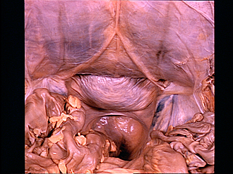
Dissection of anterolateral abdominal wall
Internal aspect of lower abdominal wall, peritoneum intact
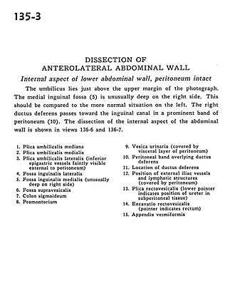
The umbilicus lies just above the upper margin of the photograph. The medial inguinal fossa (5) is unusually deep on the right side. This should be compared to the more normal situation on the left. The right ductus deferens passes toward the inguinual canal in a prominent band of peritoneum (10). The dissection of the internal aspect of the abdominal wall is shown in views 136-6 and 136-7.
- Median umbilical fold
- Medial umbilical fold
- Lateral umbilical fold (inferior epigastric vessels faintly visible external to peritoneum)
- Lateral inguinal fossa
- Medial inguinal fossa (unusually deep on right side)
- Supravesicular fossa
- Sigmoid colon
- Promontory
- Urinary bladder (covered by visceral layer of peritoneum)
- Peritoneal band overlying ductus deferens
- Location of ductus deferens
- Position of external iliac vessels and lymphatic structures (covered by peritoneum)
- Rectovesical fold (lower pointer indicates position of ureter in subperitoneal tissue)
- Rectovesical space (pointer indicates rectum)
- Appendix vermiformis