Bassett Collection of Stereoscopic Images of Human Anatomy
Dissection of anterolateral abdominal wall
Superficial vessels and nerves; Scarpa's fascia; external oblique muscles and fascia
Image #133-1
KEYWORDS: Fascia, Peripheral nervous system, Vasculature.
Creative Commons
Stanford holds the copyright to the David L. Bassett anatomical images and has assigned Creative Commons license Attribution-Share Alike 4.0 International to all of the images.

For additional information regarding use and permissions, please contact the Medical History Center.
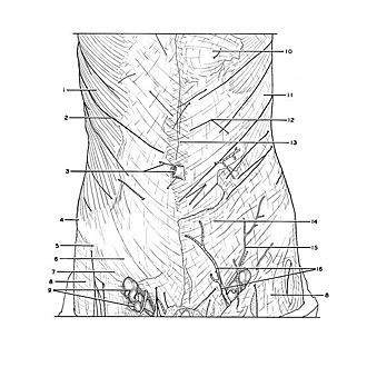
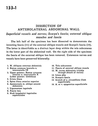
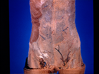
Dissection of anterolateral abdominal wall
Superficial vessels and nerves; Scarpa's fascia; external oblique muscles and fascia
The left half of the specimen has been dissected to demonstrate the investing fascia (11) of the external oblique muscle and Scarpa's fascia (14). The latter is identifiable as a distinct layer of deep within the tela subcutanea in the lower part of the abdominal wall. On the right side of the specimen the fascia of the external oblique has been removed. Cutaneous nerves and vessels have been preserved bilaterally.
- External oblique muscle
- Lateral cutaneus branch intercostal nerve IX
- Upper pointer: Anterior cutaneus branch intercostal nerve X Lower pointer: Umbilicus
- Iliac crest
- Anterior superior iliac spine
- Aponeurosis external oblique muscle
- Inguinal ligament
- Fascia lata
- Superficial inguinal lymph nodes
- Superficial fascia
- Fascia of external oblique muscle
- Rectus abdominis muscle (visible through sheath of rectus)
- Linea alba
- Scarpa's fascia
- Branch of superficial circumflex iliac artery
- Superficial epigastric artery and vein