
Bassett Collection of Stereoscopic Images of Human Anatomy
Dissection of lungs in situ
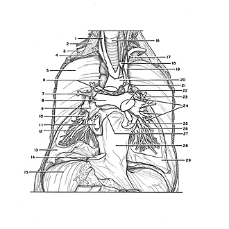
General view of vascular and bronchial distribution within lungs.
Image #124-1
KEYWORDS: Left lung, Lung, Pericardial sac, Right lung, Vasculature.
Creative Commons
Stanford holds the copyright to the David L. Bassett anatomical images and has assigned Creative Commons license Attribution-Share Alike 4.0 International to all of the images.

For additional information regarding use and permissions, please contact the Medical History Center.

Dissection of lungs in situ
General view of vascular and bronchial distribution within lungs.
The specimen is shown in a considerably more advanced stage of dissection than that in the previous view. The upper lobes of both lungs and the right middle lobe have been removed. However, the major vessels and bronchi to these parts have been preserved. Most of the heart has been removed. The ascending aorta has been cut off to expose the right pulmonary artery (8). The pericardium has been cut away except for its posterior and diaphragmatic parts.
- Right common carotid artery
- Anterior scalene muscle
- Right subclavian artery
- Vagus nerve right
- Costal pleura
- Upper pointer: Anterior mediastinal lymph node Lower pointer: Lymphatic Vessels
- Bronchus of upper right lobe
- Right pulmonary artery
- Right. superior pulmonary vein
- Bronchus of middle right lobe
- Right inferior pulmonary vein (at entrance into left atrium)
- Lateral basal segmental bronchus
- Inferior lobe right lung
- Inferior vena cava (cut off at entrance into right atrium)
- Diaphragm
- Trachea
- Pleura
- Left common carotid artery (cut off)
- Left subclavian artery
- Aortic arch
- Cardiac plexus
- Pulmonary trunk
- Left pulmonary artery
- Bronchus of upper left lobe
- Left inferior pulmonary vein
- Medial basal segmental bronchus
- Left atrium
- Pericardium
- Lower lobe left lung