Bassett Collection of Stereoscopic Images of Human Anatomy
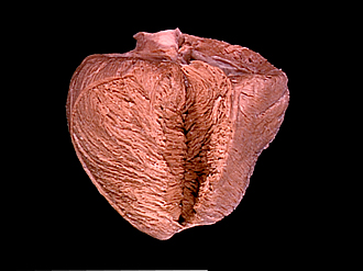
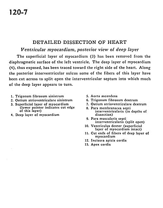
Detailed dissection of heart
Ventricular myocardium, posterior view of deep layer
Image #120-7
KEYWORDS: Heart, Left heart, Muscles and tendons, Right heart.
Creative Commons
Stanford holds the copyright to the David L. Bassett anatomical images and has assigned Creative Commons license Attribution-Share Alike 4.0 International to all of the images.

For additional information regarding use and permissions, please contact the Medical History Center.
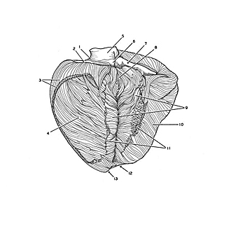
Detailed dissection of heart
Ventricular myocardium, posterior view of deep layer
The superficial layer of myocardium (3) has been removed from the diaphragmatic surface of the left ventricle. The deep layer of myocardium (4), thus exposed, has been traced toward the right side of the heart. Along the posterior interventricular sulcus some of the fibers of this layer have been cut across to split open the interventricular septum into which much of the deep layer appears to turn.
- Left fibrous trigone
- Left atrioventricular opening
- Superficial layer of myocardium (lower pointer indicates cut edge of this layer)
- Deep layer of myocardium
- Ascending aorta
- Right fibrous trigone
- Right atrioventricular opening
- Membranous part interventricular septum (in depths of dissection)
- Muscular part interventricular septum (split open)
- Right ventricle (superficial layer of myocardium intact)
- Cut ends of fibers of deep layer of myocardium
- Notch at apex of heart
- Apex of heart