Bassett Collection of Stereoscopic Images of Human Anatomy
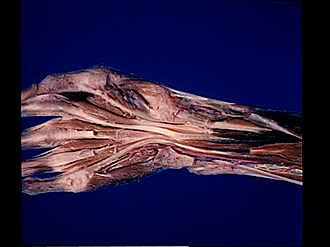
Volar aspect of right hand
Flexor digitorum sublimis muscle (continued); tendons to second and fifth fingers
Image #103-4
KEYWORDS: Fascia ligaments and tendons, Hand and fingers, Vasculature.
Creative Commons
Stanford holds the copyright to the David L. Bassett anatomical images and has assigned Creative Commons license Attribution-Share Alike 4.0 International to all of the images.

For additional information regarding use and permissions, please contact the Medical History Center.

Volar aspect of right hand
Flexor digitorum sublimis muscle (continued); tendons to second and fifth fingers
The superficial parts of the muscle (12) have been resected between the forearm and the third and fourth fingers. Remnants of the common synovial sheath (ulnar bursa) are visible over the flexor digitorum profundus (9) within the carpal canal, and distally over the lumbrical muscles (18).
- Ligament of digital sheath
- Abductor pollicis brevis muscle
- Adductor pollicis muscle (transverse head)
- Lumbrical muscle
- Flexor pollicis longus muscle (tendon of insertion)
- Abductor pollicis longus muscle (tendon of insertion)
- Flexor carpi radialis muscle (tendon of insertion)
- Radial artery
- Flexor digitorum profundus muscle (tendon to index finger, covered by synovial sheath)
- Radius (covered by periosteum)
- Flexor digitorum superficialis (deep heads, to second and fifth fingers)
- Flexor digitorum superficialis (superficial heads, resected)
- Ulnar nerve
- Ulnar artery
- Ulna (covered by periosteum)
- Flexor carpi ulnaris muscle (tendon of insertion)
- Transverse carpal ligament
- Sheath of common tendon of flexor muscles (posterior wall of opened sheath)
- Flexor digiti minimi muscle