
Bassett Collection of Stereoscopic Images of Human Anatomy
Volar aspect of forearm
Flexor pollicis longus and flexor digitorum profundus muscles
Image #98-6
KEYWORDS: Forearm, Vasculature.
Creative Commons
Stanford holds the copyright to the David L. Bassett anatomical images and has assigned Creative Commons license Attribution-Share Alike 4.0 International to all of the images.

For additional information regarding use and permissions, please contact the Medical History Center.

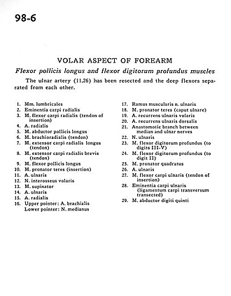
Volar aspect of forearm
Flexor pollicis longus and flexor digitorum profundus muscles
The ulnar artery (11,26)has been resected and the deep flexors separated from each other.
- Lumbrical muscles
- Radial carpal eminence
- Flexor carpi radialis muscle (tendon of insertion)
- Radial artery
- Abductor pollicis longus muscle
- Brachioradialis muscle (tendon)
- Extensor carpi radialis longus muscle (tendon)
- Extensor carpi radialis brevis muscle (tendon)
- Flexor pollicis longus muscle
- Pronator teres muscle (insertion)
- Ulnar artery
- Anterior interosseous nerve
- Supinator muscle
- Ulnar artery
- Radial artery
- Upper pointer: Brachial artery Lower pointer: Median nerve
- Muscular branch of ulnar nerve
- Pronator teres muscle (ulnar head)
- Anterior recurrent ulnar artery
- Dorsal recurrent ulnar artery
- Anastomotic branch between median and ulnar nerves
- Ulnar nerve
- Flexor digitorum profundus muscle (to digits III-V)
- Flexor digitorum profundus muscle (to digit II)
- Pronator quadratus muscle
- Ulnar artery
- Flexor carpi ulnaris muscle (tendon of insertion)
- Ulnar carpal eminence (ligamentum carpi transversum transected)
- Abductor digiti minimi muscle