Bassett Collection of Stereoscopic Images of Human Anatomy
Volar aspect of forearm
Course of ulnar nerve and artery in right forearm
Image #98-5
KEYWORDS: Forearm, Neuralnetwork, Peripheral nervous system.
Creative Commons
Stanford holds the copyright to the David L. Bassett anatomical images and has assigned Creative Commons license Attribution-Share Alike 4.0 International to all of the images.

For additional information regarding use and permissions, please contact the Medical History Center.
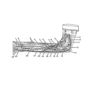
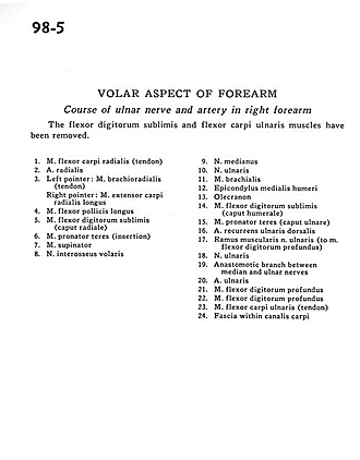
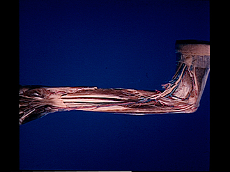
Volar aspect of forearm
Course of ulnar nerve and artery in right forearm
The flexor digitorum sublimis and flexor carpi ulnaris muscles have been removed.
- Flexor carpi radialis muscle (tendon)
- Radial artery
- Left pointer: Brachioradialis muscle (tendon) Right pointer: Extensor carpi radialis longus muscle
- Flexor pollicis longus muscle
- Flexor digitorum superficialis (radial head)
- Pronator teres muscle (insertion)
- Supinator muscle
- Anterior interosseous nerve
- Median nerve
- Ulnar nerve
- Brachialis muscle
- Medial epicondyle of humerus
- Olecranon
- Flexor digitorum superficialis (humeral head)
- Pronator teres muscle (ulnar head)
- Dorsal recurrent ulnar artery
- Muscular branch of ulnar nerve (to flexor digitorum profundus muscle)
- Ulnar nerve
- Anastomotic branch between median and ulnar nerves
- Ulnar artery
- Flexor digitorum profundus muscle
- Flexor digitorum profundus muscle
- Flexor carpi ulnaris muscle (tendon)
- Fascia within carpal tunnel