
Bassett Collection of Stereoscopic Images of Human Anatomy
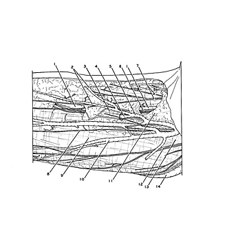
Volar aspect of forearm
Superficial veins and nerves in right cubital fossa, close-up view
Image #96-7
KEYWORDS: Fascia ligaments and tendons, Neuralnetwork, Peripheral nervous system.
Creative Commons
Stanford holds the copyright to the David L. Bassett anatomical images and has assigned Creative Commons license Attribution-Share Alike 4.0 International to all of the images.

For additional information regarding use and permissions, please contact the Medical History Center.

Volar aspect of forearm
Superficial veins and nerves in right cubital fossa, close-up view
The cubital fossa (11) is covered by a layer of fascia which blends with the lacertus fibrosus (13) and which is continuous with the antibrachial fascia (9).
- Cephalic vein
- Subcutaneous plexus of veins
- Deep lamina of superficial fascia
- Communication between superficial and deep veins
- Median cubital vein
- Lateral antebrachial cutaneous nerve
- Branches of lateral antebrachial cutaneous nerve
- Branch middle antebrachial cutaneous nerve
- Antibrachial fascia
- Median antebrachial vein
- Location of cubital fossa (covered by fascia)
- Anterior branch middle antebrachial cutaneous nerve
- Bicipital aponeurosis
- Basilic vein