Bassett Collection of Stereoscopic Images of Human Anatomy
Arm
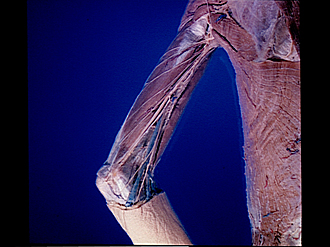
Left triceps muscle, posteromedial view
Image #96-1
KEYWORDS: Neuralnetwork, Upper arm, Vasculature.
Creative Commons
Stanford holds the copyright to the David L. Bassett anatomical images and has assigned Creative Commons license Attribution-Share Alike 4.0 International to all of the images.

For additional information regarding use and permissions, please contact the Medical History Center.


Arm
Left triceps muscle, posteromedial view
The brachial fascia has been removed. The superficial nerves which were shown in the previous view have been retained.
- Deltoid muscle
- Branch intercostobrachial nerve (displaced upward)
- Lateral brachial cutaneous axillary nerve
- Posterior brachial cutaneous nerve of radial nerve
- IntercostobrachIal nerve
- Branches of middle brachial cutaneous nerve
- Triceps brachii muscle (long head)
- Ulnar nerve
- Triceps brachii muscle (medial head)
- Upper pointer: Medial intermuscular septum Lower pointer: Brachialis muscle (covered by fascia)
- Medial epicondyle of humerus
- Infraspinatus fascia
- Teres major muscle
- Median nerve
- Latissimus dorsi muscle
- Biceps brachii muscle
- Middle antibrachial cutaneous nerve
- Basilic vein