Bassett Collection of Stereoscopic Images of Human Anatomy
Arm
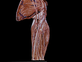
Nerve supply to left coracobrachialis, biceps and brachialis muscles, medial view
Image #95-5
KEYWORDS: Neuralnetwork, Upper arm, Vasculature.
Creative Commons
Stanford holds the copyright to the David L. Bassett anatomical images and has assigned Creative Commons license Attribution-Share Alike 4.0 International to all of the images.

For additional information regarding use and permissions, please contact the Medical History Center.


Arm
Nerve supply to left coracobrachialis, biceps and brachialis muscles, medial view
The muscles have been retracted to show the course of the musculocutaneous nerve.
- Radial nerve
- Vertebral margin of scapula
- Teres major muscle
- Thoracodorsal artery
- Latissimus dorsi muscle (tendon of insertion)
- Triceps brachii muscle
- Superior ulnar collateral artery
- Ulnar nerve
- Medial intermuscular septum
- Median nerve
- Brachial artery
- Joint of humerus
- Middle cord of brachial plexus
- Upper pointer: Musculocutaneous nerve Lower pointer: Branches of musculocutaneous nerve (to coracobrachialis muscle)
- Lateral head of median nerve
- Posterior circumflex artery of humerus
- Coracobrachialis muscle
- Medial head of median nerve
- Lymph node along course of deep brachial artery
- Biceps brachii muscle (short head)
- Biceps brachii muscle (long head)
- Branches of musculocutaneous nerve (to biceps brachii muscle)
- Muscular branch radial artery
- Radial artery (note anomalous, but not infrequent, high origin from brachial artery)
- Brachialis muscle
- Lateral antebrachial cutaneous nerve
- Branches of musculocutaneous nerve (to brachialis muscle)