Bassett Collection of Stereoscopic Images of Human Anatomy
Creative Commons
Stanford holds the copyright to the David L. Bassett anatomical images and has assigned Creative Commons license Attribution-Share Alike 4.0 International to all of the images.

For additional information regarding use and permissions, please contact the Medical History Center.

Shoulder
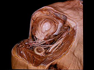
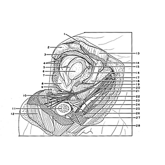
Course of right axillary nerve
The specimen shown in the previous view has been dissected to illustrate the relations of the axillary nerve. The capsule of the shoulder joint (8) has been detached from the surgical neck of the humerus. The upper part of the humerus has been cut away. Veins have been removed.
- Acromioclavicular ligament
- Acromion of scapula
- Supraspinatus muscle (tendon of insertion)
- Upper pointer: Infraspinatus muscle Lower pointer: Joint capsule of humerus
- Upper pointer: Subcoracoid bursa Lower pointer: Glenoid labrum
- Glenoid cavity
- Teres minor muscle
- Joint capsule of humerus (pointer on area of attachment to humerus)
- Branch axillary nerve (to teres minor muscle)
- Triceps brachii muscle (long head)
- Body of humerus
- Deltoid muscle
- Deltoid muscle (clavicular origin)
- Coracoid process of scapula
- Biceps brachii muscle (long head)
- Subscapularis muscle (area of insertion on lesser tubercle)
- Axillary nerve
- Biceps brachii muscle (short head)
- Radial nerve
- Subscapularis muscle
- Pectoralis minor muscle
- Musculocutaneous nerve
- Axillary artery
- Median nerve
- Posterior circumflex artery of humerus
- Circumflex scapular artery
- Pectoralis major muscle (partially resected)
- Biceps brachii muscle