Bassett Collection of Stereoscopic Images of Human Anatomy
Shoulder
Cavity of left shoulder joint, posterior view
Image #94-7
KEYWORDS: Muscles and tendons, Shoulder.
Creative Commons
Stanford holds the copyright to the David L. Bassett anatomical images and has assigned Creative Commons license Attribution-Share Alike 4.0 International to all of the images.

For additional information regarding use and permissions, please contact the Medical History Center.

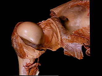
Shoulder
Cavity of left shoulder joint, posterior view
The articular capsule has been incised vertically and the humerus retracted laterally. The head of the humerus has been rotated posteriorly.
- Acromion of scapula (cut)
- Joint capsule of humerus (cut edge turned inward)
- Glenoid labrum
- Biceps brachii muscle (long head)
- Position of superior glenohumeral ligament (covered internally by articular capsule)
- Supraspinatus muscle (tendon fused with joint capsule)
- Head of humerus
- Tendon of subscapularis muscle (opening of bursa subscapularis visible)
- Middle glenohumeral ligament (covered internally by articular capsule)
- Anatomic neck of humerus
- Inferior glenohumeral ligament (covered internally by articular capsule)
- Joint capsule of humerus
- Teres minor muscle
- Joint capsule of humerus (reinforced by inferior glenohumeral ligament)
- Body of humerus
- Spinoglenoid notch
- Neck of scapula
- Glenoid cavity
- Infraspinatus muscle
- Triceps brachii muscle (long head)
- Posterior circumflex artery of humerus