Bassett Collection of Stereoscopic Images of Human Anatomy
Pectoral region and axilla
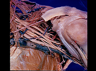
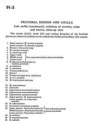
Left axilla (continued); relations of arteries, veins and nerves, close-up view
Image #91-3
KEYWORDS: Axilla, Neuralnetwork, Peripheral nervous system.
Creative Commons
Stanford holds the copyright to the David L. Bassett anatomical images and has assigned Creative Commons license Attribution-Share Alike 4.0 International to all of the images.

For additional information regarding use and permissions, please contact the Medical History Center.

Pectoral region and axilla
Left axilla (continued); relations of arteries, veins and nerves, close-up view
The trunks (5,6,7), cords (24) and various branches of the brachial plexus are shown in relation to the subclavian (9,10) and axillary (14) vessels.
- Upper pointer: levator muscle of scapula Lower pointer: Dorsal nerve of scapula
- Branch of long thoracic nerve
- Serratus anterior muscle
- Superficial transverse artery
- Upper trunk
- Middle trunk
- Lower trunk (91-4 pertain to the supraclavicular part of brachial plexus)
- Phrenic nerve (lying upon anterior scalene muscle)
- Subclavian artery
- Subclavian vein
- Subclavian trunk
- Rib I
- Tendon of origin of subclavius muscle
- Axillary artery and vein
- External intercostal muscle
- Supraspinatus muscle
- Clavicle
- Acromioclavicular ligament
- Coracoclavicular ligament
- Coracoacromial ligament
- Upper pointer: Suprascapular nerve Lower pointer: Transverse scapular artery
- Subscapular nerve
- Coracoid process of scapula
- Upper pointer: Posterior cord Middle pointer: Lateral cord Lower pointer: Middle cord (together these are infraclavicular parts of the brachial plexus)
- Axillary nerve
- Musculocutaneous nerve
- Radial nerve
- Coracobrachialis muscle
- Median nerve