Bassett Collection of Stereoscopic Images of Human Anatomy
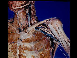
Pectoral region and axilla
Left axilla (continued); general view of neck, shoulder and axilla, clavicle intact
Image #90-6
KEYWORDS: Axilla, Pectoral region, Peripheral nervous system, Shoulder.
Creative Commons
Stanford holds the copyright to the David L. Bassett anatomical images and has assigned Creative Commons license Attribution-Share Alike 4.0 International to all of the images.

For additional information regarding use and permissions, please contact the Medical History Center.
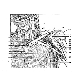

Pectoral region and axilla
Left axilla (continued); general view of neck, shoulder and axilla, clavicle intact
The left sternocleidomastoid and trapezius muscles have been removed and the deep structures of the neck dissected. The deltoid, pectoralis major, pectoralis minor and subclavius muscles have also been cut away and the axilla freed of connective tissue and lymphatic structures. The arm has been rotated slightly medially.
- Thyroid gland
- Sternocleidomastoid muscle
- Costoclavicular ligament
- Sternoclavicular joint (opened)
- Pectoralis major muscle
- Manubrium of sternum
- External intercostal muscle
- Rib II
- Internal intercostal muscle
- Internal jugular vein
- Superficial cervical artery
- Supraclavicular part of brachial plexus
- Clavicle
- Coracoclavicular ligament
- Acromioclavicular joint capsule
- Acromion
- Joint capsule of humerus
- Coracoacromial ligament
- Coracoid process of scapula
- Humerus
- Infraclavicular part of brachial plexus
- Axillary artery
- Biceps brachii muscle (long head)
- Biceps brachii muscle (short head)
- Coracobrachialis muscle
- Supreme thoracic artery
- Serratus anterior muscle
- Origin of pectoralis minor muscle