
Bassett Collection of Stereoscopic Images of Human Anatomy
Pectoral region and axilla
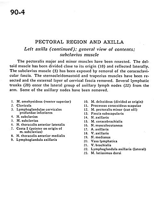
Left axilla (continued); general view of contents; subclavius muscle
Image #90-4
KEYWORDS: Axilla, Lymphatics, Peripheral nervous system.
Creative Commons
Stanford holds the copyright to the David L. Bassett anatomical images and has assigned Creative Commons license Attribution-Share Alike 4.0 International to all of the images.

For additional information regarding use and permissions, please contact the Medical History Center.

Pectoral region and axilla
Left axilla (continued); general view of contents; subclavius muscle
The pectoralis major and minor muscles have cervical been resected. The deltoid muscle has been divided close to its origin (10) and reflected laterally. The subclavius muscle (5) has been exposed by removal of the coracoclavicular fascia. The sternocleidomastoid and trapezius muscles have been resected and the external layer of cervical fascia removed. Several lymphatic trunks (20) enter the lateral group of axillary lymph nodes (22) from the arm. Some of the axillary nodes have been removed.
- Omohyoid muscle (superior belly)
- Clavicle
- Inferior deep cervical lymph nodes
- Subclavian nerve
- Subclavius muscle
- Anterior lateral thoracic nerve
- Rib I (pointer on origin of subclavius muscle)
- Anterior medial thoracic nerve
- Axillary lymph node
- Deltoid muscle (divided at origin)
- Coracoid process of scapula
- Pectoralis minor muscle (cut off)
- Subscapular fascia
- Axillary nerve
- Coracobrachialis muscle
- Musculocutaneous nerve
- Axillary artery
- Axillary vein
- Median nerve
- Lymph vessel
- Brachial vein
- Axillary lymph node (lateral)
- Latissimus dorsi muscle