Bassett Collection of Stereoscopic Images of Human Anatomy
Exploration of the brain from its superior aspect
Close-up view of anterior part of lateral ventricle and interventricular foramen
Image #9-4
KEYWORDS: Meninges, Ventricules.
Creative Commons
Stanford holds the copyright to the David L. Bassett anatomical images and has assigned Creative Commons license Attribution-Share Alike 4.0 International to all of the images.

For additional information regarding use and permissions, please contact the Medical History Center.
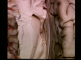
Exploration of the brain from its superior aspect
Close-up view of anterior part of lateral ventricle and interventricular foramen
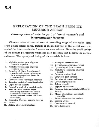
Close-up view of central area of preceding stage of dissection seen from a more lateral angle. Details of the medial wall of the lateral ventricle and of the interventricular foramen are now evident. Note the small cavity of the septum pellucidum which has been cut open just beneath the corpus callosum. The ependymal lining of the ventricle is intact.
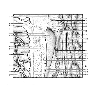
- Medullary substance of superior frontal gyrus
- Medullary substance of inferior frontal gyrus
- Junction of fibers from internal capsule and corpus callosum to form corona radiata (seen in cross section)
- Anterior horn lateral ventricle
- Superior occipitofrontal fasciculus
- Head of caudate nucleus
- Frontal branch of middle cerebral artery
- Area of fibers derived from medullary substance of insula, external capsule and internal capsule (fibers course in various directions)
- Emerging fibers of internal capsule
- Insula
- Artery of precentral sulcus
- Artery of central sulcus
- Transverse temporal gyrus
- Cingulate gyrus (cut across)
- Medullary substance of cingulate gyrus
- Genu corpus callosum
- Cingulum (cut across)
- Cut edge of corpus callosum
- Cavum septum pellucidum
- Cingulate sulcus
- Septum pellucidum
- Interventricular foramen (of Monro)
- Fornix
- Choroid plexus in lateral ventricle
- Anterior thalamic tubercle
- Lamina affixa
- Caudate nucleus (tail)
- Stria terminalis