Bassett Collection of Stereoscopic Images of Human Anatomy
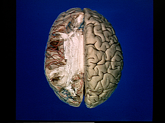
Exploration of the brain from its superior aspect
Lateral fissure and insula
Image #9-1
KEYWORDS: Brain, Telencephalon, Temporal lobe, Vasculature.
Creative Commons
Stanford holds the copyright to the David L. Bassett anatomical images and has assigned Creative Commons license Attribution-Share Alike 4.0 International to all of the images.

For additional information regarding use and permissions, please contact the Medical History Center.
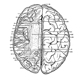

Exploration of the brain from its superior aspect
Lateral fissure and insula
The lateral fissure has now been extensively exposed from above by the removal of the frontal and parietal opercula. The superior longitudinal fasciculus (4) has been cut away to reveal the insula. The principal cortical branches of the middle cerebral artery lie within the lateral fissure. The portion of the temporal lobe facing the fissure displays transversely oriented gyri (of Heschl) wherein the fibers of the central auditory pathway terminate.
- Medullary substance of superior frontal gyrus
- Superior cerebral vein is in superior frontal sulcus
- Cingulum (cut off)
- Anterior continuation of superior longitudinal fasciculus
- Frontal branches of orbitofrontal artery
- Insula
- Artery of precentral sulcus
- Artery of central sulcus (Note cut end of great anastomotic vein in meninges close to cut end of this artery)
- Transverse temporal gyrus (of Heschl)
- Sulcus circularis Note: this sulcus surrounds the insula in the depths of the lateral fissure
- Anterior and posterior parietal arteries
- Posterior portion of superior longitudinal fasciculus (cut off)
- Geniculocalcarine tract and inferior longitudinal fasciculus joining occipital part internal capsule
- Occipital part internal capsule and occipital part of radiations of corpus callosum
- Branch of posterior cerebral artery in depths of parieto-occipital fissure
- Branch of posterior cerebral artery in calcarine fissure (exposed by removing cuneus)
- Medullary substance of cuneus
- Frontal pole
- Superior frontal gyrus
- Cingulate gyrus (cut across)
- Central sulcus (Rolandic)
- Corona radiata
- Medial and lateral longitudinal striae