Bassett Collection of Stereoscopic Images of Human Anatomy
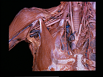
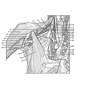
Pectoral region and axilla
Right pectoralis minor muscle
Image #89-4
KEYWORDS: Pectoral region, Peripheral nervous system, Vasculature.
Creative Commons
Stanford holds the copyright to the David L. Bassett anatomical images and has assigned Creative Commons license Attribution-Share Alike 4.0 International to all of the images.

For additional information regarding use and permissions, please contact the Medical History Center.

Pectoral region and axilla
Right pectoralis minor muscle
The relations of the pectoralis minor muscle to the axillary artery and brachial plexus can be seen. The subclavian and axillary veins have been removed.
- Supraclavicular part of brachial plexus (pointer on upper trunk)
- Trapezius muscle
- Omohyoid muscle (cut off)
- Clavicle (cut off)
- Deltoid muscle
- Coracoid process of scapula
- Coracobrachialis muscle
- Musculocutaneous nerve
- Cephalic vein
- Pectoralis major muscle (cut off)
- Median nerve
- Brachial veins
- Axilla
- Pectoralis minor muscle
- Latissimus dorsi muscle
- Superficial cervical artery
- Common carotid artery
- Superficial transverse artery
- Transverse scapular artery
- Subclavian artery
- Anterior scalene muscle
- Trachea
- Left pointer: Lateral cord of brachial plexus Right pointer: Axillary artery
- Brachiocephalic artery
- Brachiocephalic veins
- Ascending aorta
- Internal mammary artery
- Rib II