
Bassett Collection of Stereoscopic Images of Human Anatomy
Pectoral region and axilla
Right pectoralis major and deltoid muscles, anterior view
Image #89-2
KEYWORDS: Forearm, Pectoral region, Vasculature.
Creative Commons
Stanford holds the copyright to the David L. Bassett anatomical images and has assigned Creative Commons license Attribution-Share Alike 4.0 International to all of the images.

For additional information regarding use and permissions, please contact the Medical History Center.
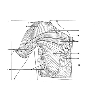

Pectoral region and axilla
Right pectoralis major and deltoid muscles, anterior view
The deep fascia has been removed. A well-developed sternalis muscle (9) is present. Later dissection revealed that this variant was not bilateral.
- Acromion
- Deltoid muscle
- Biceps brachii muscle (upper pointer, short head; lower pointer, long head)
- Serratus anterior muscle
- Clavicle
- Sternocleidomastoid muscle
- Clavicular part pectoralis major muscle
- Sternocostal part pectoralis major muscle
- Sternalis muscle
- Sternum