
Bassett Collection of Stereoscopic Images of Human Anatomy
Hyoid bone and framework of larynx
Cricoid and arytenoid cartilages, right anterolateral view
Image #84-7
KEYWORDS: Bones cartilage joints.
Creative Commons
Stanford holds the copyright to the David L. Bassett anatomical images and has assigned Creative Commons license Attribution-Share Alike 4.0 International to all of the images.

For additional information regarding use and permissions, please contact the Medical History Center.

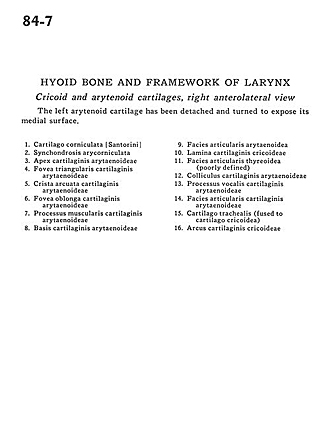
Hyoid bone and framework of larynx
Cricoid and arytenoid cartilages, right anterolateral view
The left arytenoid cartilage has been detached and turned to expose its medial surface.
- Corniculate cartilage
- Arycorniculate synchondrosis
- Apex of arytenoid cartilage
- Triangular fovea of arytenoid cartilage
- Arcuate crest of arytenoid cartilage
- Fovea oblonga arytenoid cartilage
- Muscular process of arytenoid cartilage
- Base of arytenoid cartilage
- Articular surface arytenoid
- Lamina of cricoid cartilage
- Articular surface thyroid (poorly defined)
- Arytenoid cartilaginous neck
- Vocal process of arytenoid cartilage
- Articular surface arytenoid cartilage
- Tracheal cartilage (fused to cricoid cartilage)
- Arch of cricoid cartilage