
Bassett Collection of Stereoscopic Images of Human Anatomy
Dissection of head and neck from a posterior approach
Larynx; blood vessels and nerves
Image #83-3
KEYWORDS: Peripheral nervous system, Throat, Vasculature.
Creative Commons
Stanford holds the copyright to the David L. Bassett anatomical images and has assigned Creative Commons license Attribution-Share Alike 4.0 International to all of the images.

For additional information regarding use and permissions, please contact the Medical History Center.
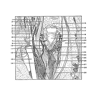
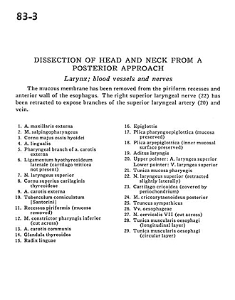
Dissection of head and neck from a posterior approach
Larynx; blood vessels and nerves
The mucous membrane has been removed from the piriform recesses and anterior wall of the esophagus. The right superior laryngeal nerve (22) has been retracted to expose branches of the superior laryngeal artery (20) and vein.
- External maxillary artery
- Salpingopharyngeus muscle
- Greater horn hyoid bone
- Lingual artery
- Pharyngeal branch of external carotid artery
- Lateral thyrohyoid ligament (Triticeal cartilage not present)
- Superior laryngeal nerve
- Superior horn thyroid cartilage
- External carotid artery
- Corniculate tubercle
- Piriform recess (mucosa removed)
- Inferior pharyngeal constrictor muscle (cut across)
- Common carotid artery
- Thyroid gland
- Root of tongue
- Epiglottis
- Pharyngoepiglottic fold (mucosa preserved)
- Aryepiglottic fold (inner mucosal surface preserved)
- Laryngeal ventricle
- Upper pointer: Superior laryngeal artery Lower pointer: Superior laryngeal vein
- Mucosa of pharynx (tunica)
- Superior laryngeal nerve (retracted slightly laterally)
- Cricoid cartilage (covered by perichondrium)
- Posterior cricoarytenoid muscle
- Sympathetic trunk
- Esophageal veins
- Cervical nerve VII (cut across)
- Muscular coat of esophagus (longitudinal layer)
- Muscular coat of esophagus (circular layer)