Bassett Collection of Stereoscopic Images of Human Anatomy
Dissection of head and neck from a posterior approach
Muscles originating from styloid process
Image #82-5
KEYWORDS:
Creative Commons
Stanford holds the copyright to the David L. Bassett anatomical images and has assigned Creative Commons license Attribution-Share Alike 4.0 International to all of the images.

For additional information regarding use and permissions, please contact the Medical History Center.
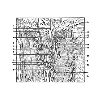
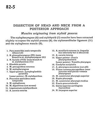
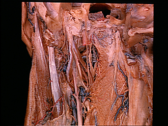
Dissection of head and neck from a posterior approach
Muscles originating from styloid process
The stylopharyngeus (6) and stylohyoid (7) muscles have been retracted slightly to expose the styloid process (8), the stylomandibular ligament (11) and the styloglossus muscle (9).
- Mastoid part of temporal bone (dissected)
- Glossopharyngeal nerve (IX) (note branch to stylopharyngeus muscle (6))
- Facial nerve (VII) (note branch to stylohyoid muscle (7))
- Parotid gland
- External pterygoid muscle
- Stylopharyngeus muscle
- Upper pointer: Lymph node parotid Lower pointer: Stylohyoid muscle
- Styloid process temporal bone
- Styloglossus muscle
- Digastric muscle (cut across)
- Stylohyoid ligament
- External carotid artery
- External maxillary artery (lingual artery lies inferiorly but is obscured)
- Internal carotid artery
- Upper pointer: Pharyngobasilar fascia Lower pointer: Pharyngeal tonsil
- Occipital condyle
- Ascending pharyngeal artery (cut off)
- Pterygoid venous plexus (near junction with pharyngeal venous plexus)
- Superior pharyngeal constrictor muscle
- Pharyngeal raphe
- Middle pharyngeal constrictor muscle
- Prevertebral fascia
- Superior horn thyroid cartilage
- Superior laryngeal nerve