Bassett Collection of Stereoscopic Images of Human Anatomy
Dissection of head and neck from a posterior approach
Middle constrictor muscle of pharynx; pharyngeal raphe; submucosal plexus of esophageal veins
Image #82-4
KEYWORDS: Muscles and tendons, Pharynx, Throat, Vasculature.
Creative Commons
Stanford holds the copyright to the David L. Bassett anatomical images and has assigned Creative Commons license Attribution-Share Alike 4.0 International to all of the images.

For additional information regarding use and permissions, please contact the Medical History Center.
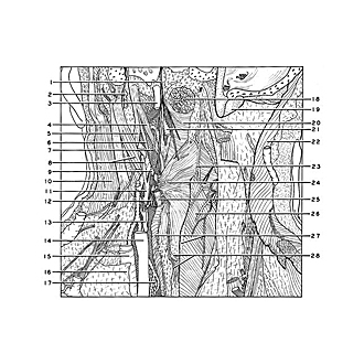
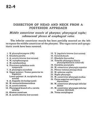
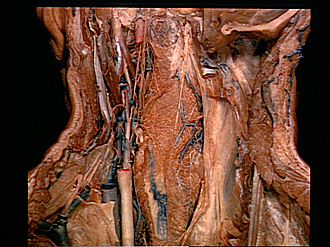
Dissection of head and neck from a posterior approach
Middle constrictor muscle of pharynx; pharyngeal raphe; submucosal plexus of esophageal veins
The inferior constrictor muscle has been partially resected on the left to expose the middle constrictor of the pharynx. The vagus nerve and sympathetic trunk have been removed.
- Glossopharyngeal nerve (IX)
- Parotid gland
- Internal carotid artery (cut across)
- Stylopharyngeus muscle
- Stylohyoid muscle
- Styloid process temporal bone
- Ascending pharyngeal artery
- Upper pointer: Posterior belly of digastric muscle Lower pointer: Occipital artery (cut across)
- Lingual artery (in background)
- Sternocleidomastoid muscle
- External carotid artery
- Pharyngeal branch of external carotid artery
- Carotid body
- Internal carotid artery (cut across)
- Internal jugular vein (cut across)
- Carotid sheath
- Thyroid gland
- Pharyngeal tonsil (pharyngobasilar fascia removed)
- Occipital condyle
- Superior pharyngeal constrictor muscle
- Pterygoid venous plexus
- Splenius capitis muscle
- Pharyngeal raphe
- Middle pharyngeal constrictor muscle
- Superior horn thyroid cartilage
- Inferior pharyngeal constrictor muscle right
- Inferior pharyngeal constrictor muscle left (divided)
- Esophageal vein