Bassett Collection of Stereoscopic Images of Human Anatomy
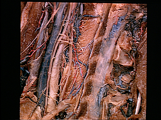
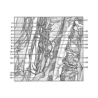
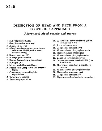
Dissection of head and neck from a posterior approach
Pharyngeal blood vessels and nerves
Image #81-6
KEYWORDS: Peripheral nervous system, Pharynx, Throat, Vasculature.
Creative Commons
Stanford holds the copyright to the David L. Bassett anatomical images and has assigned Creative Commons license Attribution-Share Alike 4.0 International to all of the images.

For additional information regarding use and permissions, please contact the Medical History Center.
Dissection of head and neck from a posterior approach
Pharyngeal blood vessels and nerves
- Hypoglossal nerve (XII)
- Nodose ganglion of vagus nerve
- Internal carotid artery
- Gray rami communicantes (to cervical nerve IV-V, which have been cut away)
- Accessory nerve (XI)
- Superior laryngeal nerve
- Descending branch hypoglossal nerve
- Vagus nerve (X)
- Sternocleidomastoid muscle
- Superficial fascia (deep lamina of external layer)
- Superior horn thyroid cartilage
- Internal jugular vein
- Sympathetic trunk
- Gray rami communicantes (to cervical nerve IV-V)
- Common carotid artery
- Ganglion cervical nerve VI
- Superior pharyngeal constrictor muscle
- Pharyngeal venous plexus
- Middle pharyngeal constrictor muscle
- Superior cervical ganglion
- Body cervical vertebra III (cut in midline)
- Pharyngeal branch of external maxillary artery
- Inferior pharyngeal constrictor muscle
- Pharyngeal plexus vagus nerve
- Ganglion cervical nerve V
- Posterior longitudinal ligament