Bassett Collection of Stereoscopic Images of Human Anatomy
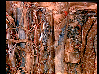
Dissection of head and neck from a posterior approach
Nerve supply to longus capitis, longus colli and rectus capitis anterior muscles
Image #81-2
KEYWORDS: Muscles and tendons, Peripheral nervous system.
Creative Commons
Stanford holds the copyright to the David L. Bassett anatomical images and has assigned Creative Commons license Attribution-Share Alike 4.0 International to all of the images.

For additional information regarding use and permissions, please contact the Medical History Center.
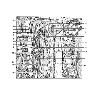

Dissection of head and neck from a posterior approach
Nerve supply to longus capitis, longus colli and rectus capitis anterior muscles
The first three cervical nerves have been retracted laterally so that their muscular branches are visible. The muscles have been elevated.
- Hypoglossal nerve (XII)
- Internal jugular vein
- Accessory nerve (XI)
- Vagus nerve (X)
- Internal carotid nerve (from superior cervical ganglion)
- Anterior branch of cervical nerve I
- Loop between first and second cervical nerves (note communications with hypoglossal nerve)
- Gray ramus communicans (to cervical nerve I)
- Anterior branch of cervical nerve II
- Upper pointer: Ganglion cervical nerve III Lower pointer: Posterior branch cervical nerve III
- Gray rami communicantes (to cervical nerves II-III)
- Superior cervical ganglion
- Internal carotid artery
- Longus capitis muscle
- Anterior arch atlas
- Dens axis
- Anterior rectus capitis muscle
- Muscular branches of cervical nerve I
- Muscular branches of cervical nerve II
- Intervertebral disc (C. II-III)
- Muscular branches of cervical nerve III
- Longus colli muscle