
Bassett Collection of Stereoscopic Images of Human Anatomy
Exploration of the brain from its superior aspect
Cingulum
Image #8-5
KEYWORDS: Brain, Parietal lobe, Telencephalon, Overview.
Creative Commons
Stanford holds the copyright to the David L. Bassett anatomical images and has assigned Creative Commons license Attribution-Share Alike 4.0 International to all of the images.

For additional information regarding use and permissions, please contact the Medical History Center.

Exploration of the brain from its superior aspect
Cingulum
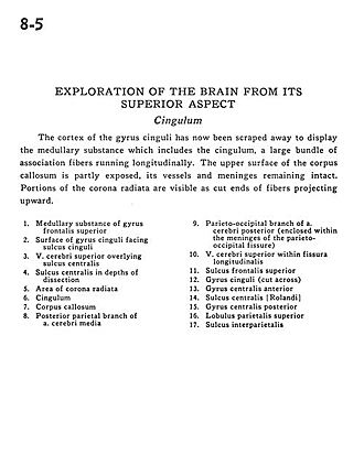
The cortex of the gyrus cinguli has now been scraped away to display the medullary substance which includes the cingulum, a large bundle of association fibers running longitudinally. The upper surface of the corpus callosum is partly exposed, its vessels and meninges remaining intact. Portions of the corona radiata are visible as cut ends of fibers projecting upward.
- Medullary substance of superior frontal gyrus
- Surface of cingulate gyrus facing cingulate sulcus
- Superior cerebral vein overlying central sulcus
- Central sulcus in depths of dissection
- Area of corona radiata
- Cingulum
- Corpus callosum
- Posterior parietal branch of middle cerebral artery
- Parieto-occipital branch of posterior cerebral artery (enclosed within the meninges of the parieto-occipital fissure)
- Superior cerebral vein within longitudinal fissure
- Superior frontal sulcus
- Cingulate gyrus (cut across)
- Precentral gyrus
- Central sulcus (Rolandic)
- Postcentral gyrus
- Superior parietal lobule
- Interparietal sulcus