Bassett Collection of Stereoscopic Images of Human Anatomy
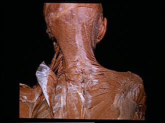
Dissection of head and neck from a posterior approach
Left splenius capitis and cervicis muscles; origin of levator scapulae muscle
Image #79-1
KEYWORDS: Connective tissue, Muscles and tendons, Fascia and connective tissue.
Creative Commons
Stanford holds the copyright to the David L. Bassett anatomical images and has assigned Creative Commons license Attribution-Share Alike 4.0 International to all of the images.

For additional information regarding use and permissions, please contact the Medical History Center.
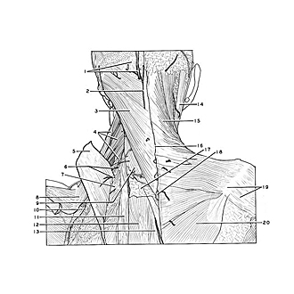
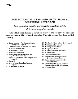
Dissection of head and neck from a posterior approach
Left splenius capitis and cervicis muscles; origin of levator scapulae muscle
The left rhomboid muscles have been removed and the serratus posterior superior muscle (5) reflected laterally. The left scapula has been pulled laterally.
- Upper pointer: Fascia covering semispinalis capitis muscle Lower pointer: Greater occipital nerve
- Third occipital nerve
- Splenius capitis muscle
- Levator scapulae muscle
- Posterior superior serratus muscle (reflected laterally)
- Nerve supply to posterior superior serratus muscle
- Rib II
- Area of insertion of levator scapulae muscle
- Splenius cervicis muscle
- Rhomboid minor muscle (cut across)
- Iliocostalis cervicis muscle
- Longissimus capitis muscle
- Dorsalis muscle
- Sternocleidomastoid muscle
- Trapezius muscle
- Nuchal ligament
- Band of lumbodorsal fascia (removed elsewhere)
- Spinous process thoracic vertebrae II
- Upper pointer: Spine of scapula Lower pointer: Deltoid muscle
- Trapezius muscle