Bassett Collection of Stereoscopic Images of Human Anatomy
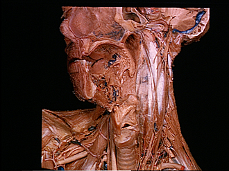
Dissection of anterior and lateral regions of neck
Cervical sympathetic trunk; vagus nerve; prevertebral muscles, anterior view
Image #78-2
KEYWORDS: Face, Mouth, Muscles and tendons, Peripheral nervous system, Pharynx, Throat.
Creative Commons
Stanford holds the copyright to the David L. Bassett anatomical images and has assigned Creative Commons license Attribution-Share Alike 4.0 International to all of the images.

For additional information regarding use and permissions, please contact the Medical History Center.
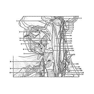

Dissection of anterior and lateral regions of neck
Cervical sympathetic trunk; vagus nerve; prevertebral muscles, anterior view
The tongue has been removed and the pharynx and palate have been sectioned near the midline. The prevertebral fascia has been cut away to the left of the midline.
- Upper pointer: Sphenoid ridge Lower pointer: Nasal part pharynx
- Soft palate (cut in midline)
- Palatine tonsil
- Uvula
- Oral part pharynx
- Geniohyoid muscle
- Body hyoid bone (cut in midline)
- Thyroid cartilage (cut across)
- Anterior scalene muscle
- Upper pointer: Vagus nerve (X) Lower pointer: Common carotid artery
- Upper pointer: Internal carotid artery (in carotid canal) Lower pointer: Jugular foramen (opened)
- Lateral rectus capitis muscle
- Longus capitis muscle
- Accessory nerve (XI)
- Upper pointer: Nodose ganglion Lower pointer: Hypoglossal nerve (XII)
- Glossopharyngeal nerve (IX)
- Superior cervical ganglion
- Superior cardiac branch vagus nerve
- Anterior longitudinal ligament
- Vagus nerve (X)
- Longus colli muscle
- Prevertebral fascia
- Sympathetic trunk
- Epiglottis
- Origins of anterior scalene muscle
- Cervical nerve VI
- Middle scalene muscle
- Posterior scalene muscle
- Cervical nerve VIII
- Inferior cervical ganglion
- Costocervical trunk