Bassett Collection of Stereoscopic Images of Human Anatomy
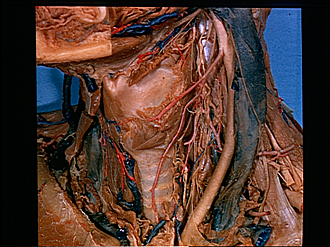
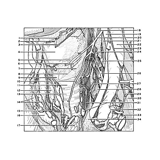
Dissection of anterior and lateral regions of neck
Relations of larynx, anterolateral view
Image #77-1
KEYWORDS: Throat.
Creative Commons
Stanford holds the copyright to the David L. Bassett anatomical images and has assigned Creative Commons license Attribution-Share Alike 4.0 International to all of the images.

For additional information regarding use and permissions, please contact the Medical History Center.
Dissection of anterior and lateral regions of neck
Relations of larynx, anterolateral view
The infrahyoid muscles have been cut away. The left lobe of the thyroid gland has been removed. The left common carotid artery, internal jugular vein and vagus nerve have been retracted laterally.
- Omohyoid muscle (cut across)
- Sternohyoid muscle (cut across)
- Thyrohyoid muscle (divided and elevated)
- Thyroid cartilage
- Cut ends of fibers of thyrohyoid muscle
- Sternothyroid muscle (cut across)
- Inferior pharyngeal constrictor muscle
- Cricothyroid muscle
- Cricoid cartilage
- Anterior branch superior thyroid artery
- Trachea
- Right lobe thyroid gland
- Isthmus of thyroid gland (cut across)
- Brachiocephalic trunk right (duplicated)
- Sternothyroid muscle (cut across)
- Anterior jugular vein
- Thymus
- Superior cardiac branch vagus nerve
- Common carotid artery (retracted laterally)
- Superior thyroid artery
- Vagus nerve (X) (retracted laterally)
- Internal jugular vein (retracted laterally)
- Descending branch hypoglossal nerve
- Sympathetic trunk
- Middle cervical ganglion
- Thyrocervical trunk
- Middle cardiac nerve
- Inferior cervical ganglion
- Phrenic nerve
- Anterior scalene muscle
- Upper pointer: Recurrent laryngeal nerve Lower pointer: Paratracheal lymph node
- Termination of lymphatic ducts in internal jugular vein
- Subclavian vein
- Fascia extending into mediastinum from carotid sheath