Bassett Collection of Stereoscopic Images of Human Anatomy
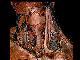
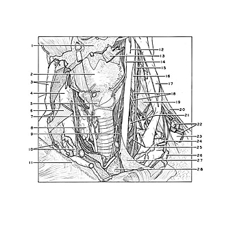
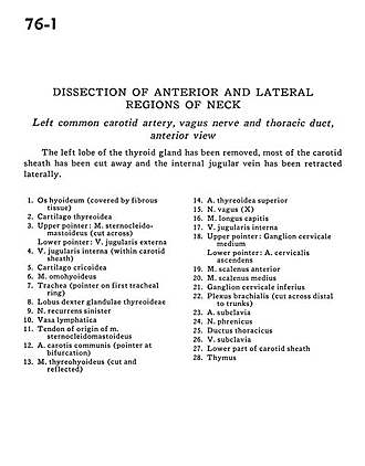
Dissection of anterior and lateral regions of neck
Left common carotid artery, vagus nerve and thoracic duct, anterior view
Image #76-1
KEYWORDS: Lymphatics, Peripheral nervous system, Vasculature.
Creative Commons
Stanford holds the copyright to the David L. Bassett anatomical images and has assigned Creative Commons license Attribution-Share Alike 4.0 International to all of the images.

For additional information regarding use and permissions, please contact the Medical History Center.
Dissection of anterior and lateral regions of neck
Left common carotid artery, vagus nerve and thoracic duct, anterior view
The left lobe of the thyroid gland has been removed, most of the carotid sheath has been cut away and the internal jugular vein has been retracted laterally.
- Hyoid bone (covered by fibrous tissue)
- Thyroid cartilage
- Upper pointer: Sternocleidomastoid muscle (cut across) Lower pointer: External jugular vein
- Internal jugular vein (within carotid sheath)
- Cricoid cartilage
- Omohyoid muscle
- Trachea (pointer on first tracheal ring)
- Right lobe of thyroid gland
- Recurrent laryngeal nerve left
- Lymph vessels
- Tendon of origin of sternocleidomastoid muscle
- Common carotid artery (pointer at bifurcation)
- Thyrohyoid muscle (cut and reflected)
- Superior thyroid artery
- Vagus nerve (X)
- Longus capitis muscle
- Internal jugular vein
- Upper pointer: Middle cervical ganglion Lower pointer: Ascending cervical artery
- Anterior scalene muscle
- Middle scalene muscle
- Inferior cervical ganglion
- Brachial plexus (cut across distal to trunks)
- Subclavian artery
- Phrenic nerve
- Thoracic duct
- Subclavian vein
- Lower part of carotid sheath
- Thymus