Bassett Collection of Stereoscopic Images of Human Anatomy
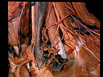
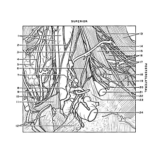
Dissection of anterior and lateral regions of neck
Relation of subclavian artery and vein to scalene muscles and brachial plexus, lateral view
Image #75-7
KEYWORDS: Muscles and tendons, Vasculature.
Creative Commons
Stanford holds the copyright to the David L. Bassett anatomical images and has assigned Creative Commons license Attribution-Share Alike 4.0 International to all of the images.

For additional information regarding use and permissions, please contact the Medical History Center.
Dissection of anterior and lateral regions of neck
Relation of subclavian artery and vein to scalene muscles and brachial plexus, lateral view
- Upper pointer: Internal jugular vein Lower pointer: Common carotid artery
- Ansa hypoglossi
- Ascending cervical artery
- Anterior branch superior thyroid artery
- Thyroid gland
- Phrenic nerve
- Transverse scapular artery
- Lymphatic trunks terminating in internal jugular vein
- Trachea
- Inferior thyroid vein left
- Sternohyoid muscle
- Upper pointer: Sternoclavicular joint capsule Lower pointer: Costoclavicular ligament
- Upper pointer: Nerves to levator scapulae muscle Lower pointer: Levator scapulae muscle
- Superficial cervical artery (posterior end displaced)
- Middle scalene muscle
- Upper pointer: Posterior scalene muscle Lower pointer: Transversa colli artery
- Long thoracic nerve (plexiform)
- Brachial plexus (pointer on divisions of superior trunk)
- Brachial plexus (pointer on divisions of middle trunk)
- Brachial plexus (pointer on divisions of inferior trunk)
- Subclavian artery
- Serratus anterior muscle
- Anterior scalene muscle (pointer near origin of muscle from first rib)
- Subclavian vein