
Bassett Collection of Stereoscopic Images of Human Anatomy
Dissection of anterior and lateral regions of neck
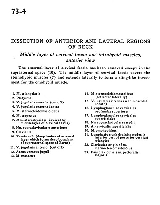
Middle layer of cervical fascia and infrahyoid muscles, anterior view
Image #73-4
KEYWORDS: Fascia and connective tissue, Muscles and tendons, Throat.
Creative Commons
Stanford holds the copyright to the David L. Bassett anatomical images and has assigned Creative Commons license Attribution-Share Alike 4.0 International to all of the images.

For additional information regarding use and permissions, please contact the Medical History Center.

Dissection of anterior and lateral regions of neck
Middle layer of cervical fascia and infrahyoid muscles, anterior view
The external layer of cervical fascia has been removed except in suprasternal space (10). The middle layer of cervical fascia covers the sternohyoid muscles (7) and extends laterally to form a sling-like investment for the omohyoid muscle.
- Depressor anguli oris muscle
- Platysma
- Anterior jugular vein (cut off)
- External jugular vein right
- Sternocleidomastoid muscles
- Trapezius muscles
- Sternohyoid muscles (covered by middle layer of cervical fascia)
- Anterior supraclavicular nerves
- Clavicle
- Superficial fascia (deep lamina of external layer which forms deep boundary of suprasternal space of Bums)
- Anterior jugular vein (cut off)
- Jugular venous arch
- Masseter muscle
- Sternocleidomastoid muscle (reflected laterally)
- Internal jugular vein (within carotid sheath)
- Superior deep cervical lymph nodes
- Superficial cervical lymph nodes
- Middle supraclavicular nerves
- Superficial cervical artery
- Omohyoid muscle
- Lymphatic trunk draining nodes in inferior part of posterior cervical triangle
- Clavicular origin of sternocleidomastoid muscle
- Clavicular part pectoralis major muscle